Raghav Narasimhan Talks about IBMS-Corneal Biomechanics
Corneal Biomechanics
Overview
Introduction
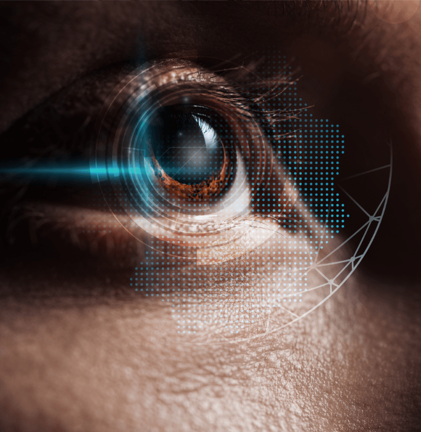
The human eye is a complex biomechanical structure in which the strength of its front surface, the cornea, is crucial for maintaining clear vision. Subtle biomechanical changes can precede clinically detectable disease, such as ectasia after laser refractive surgery or progressive bulging in keratoconus. Research at IBMS uses sophisticated tools such as viscoelastic models and finite element analysis to study how corneal mechanics affect visual quality and to predict surgical/therapeutic outcomes.
Why This Research Matters
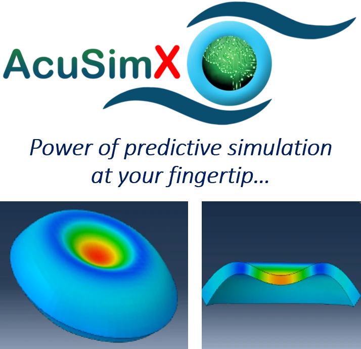
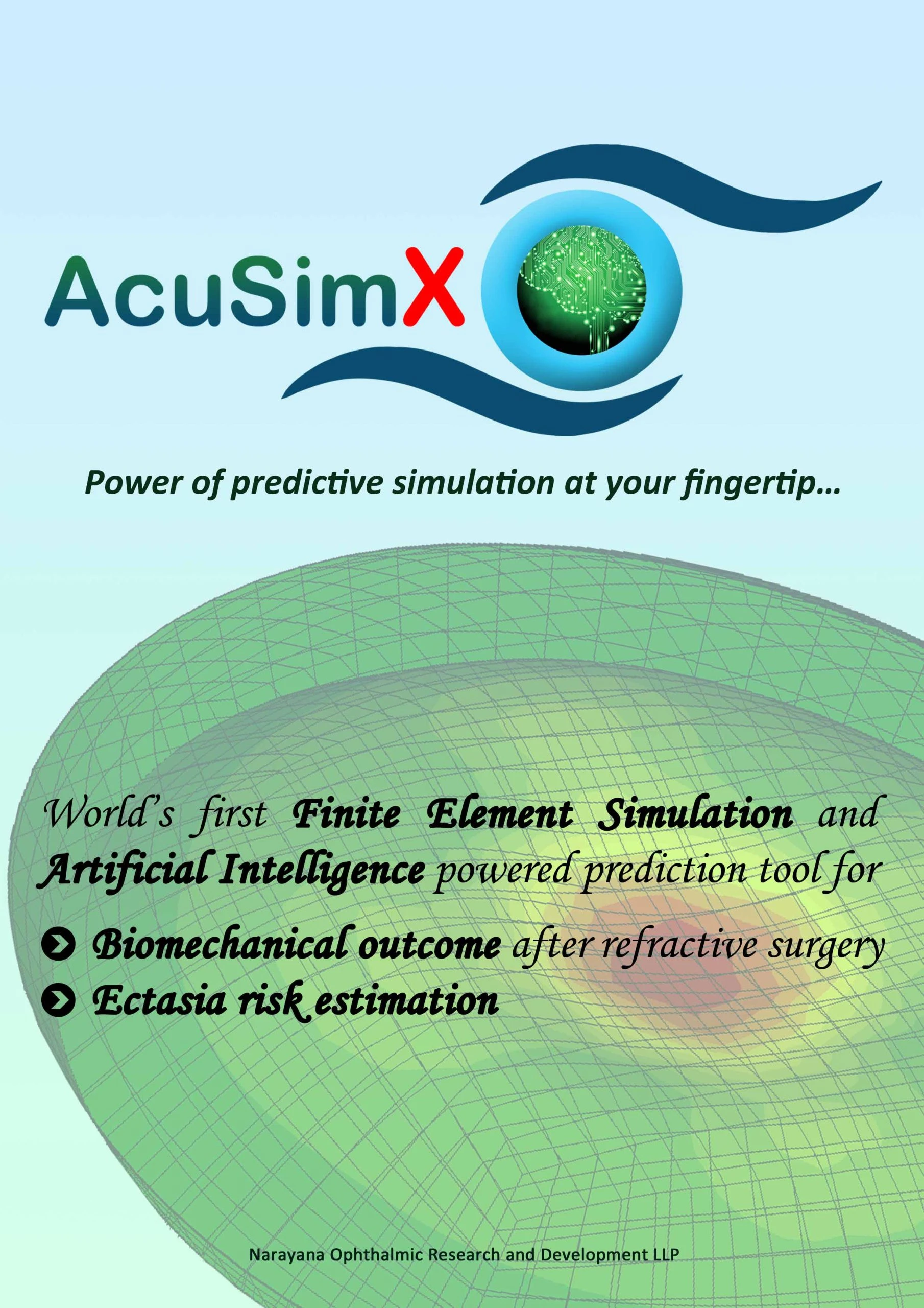
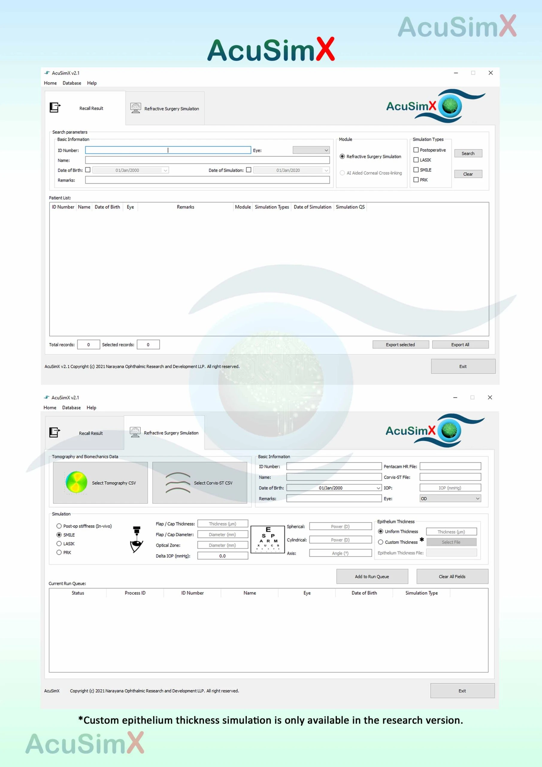
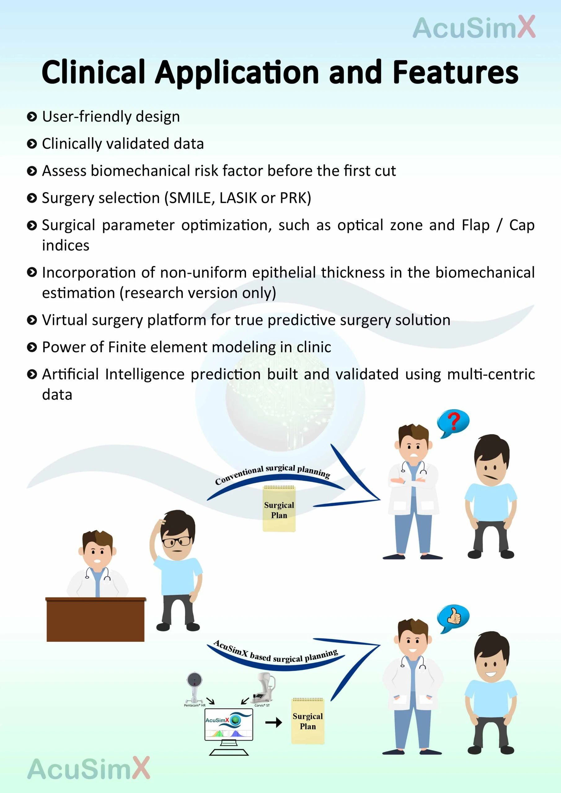
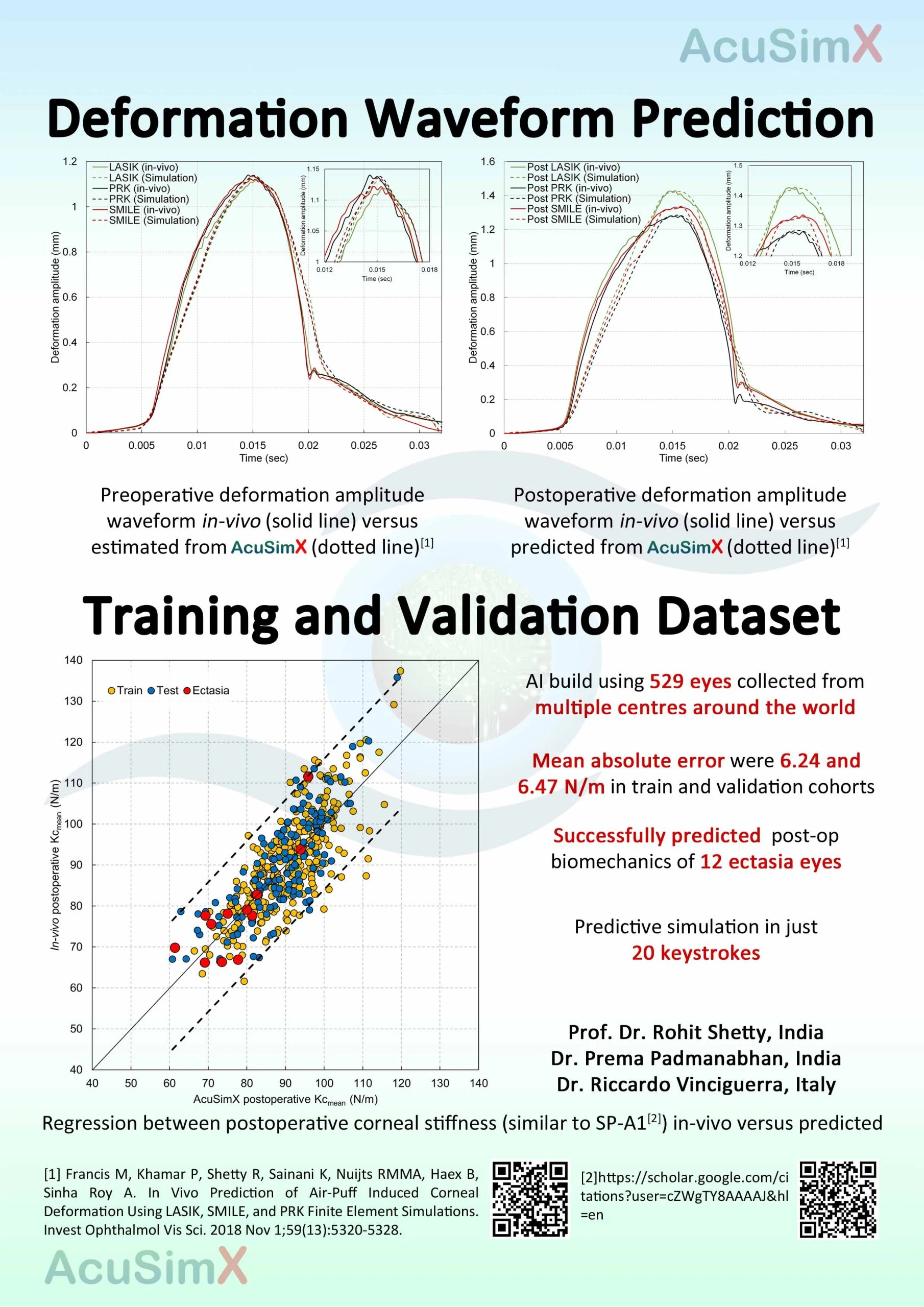
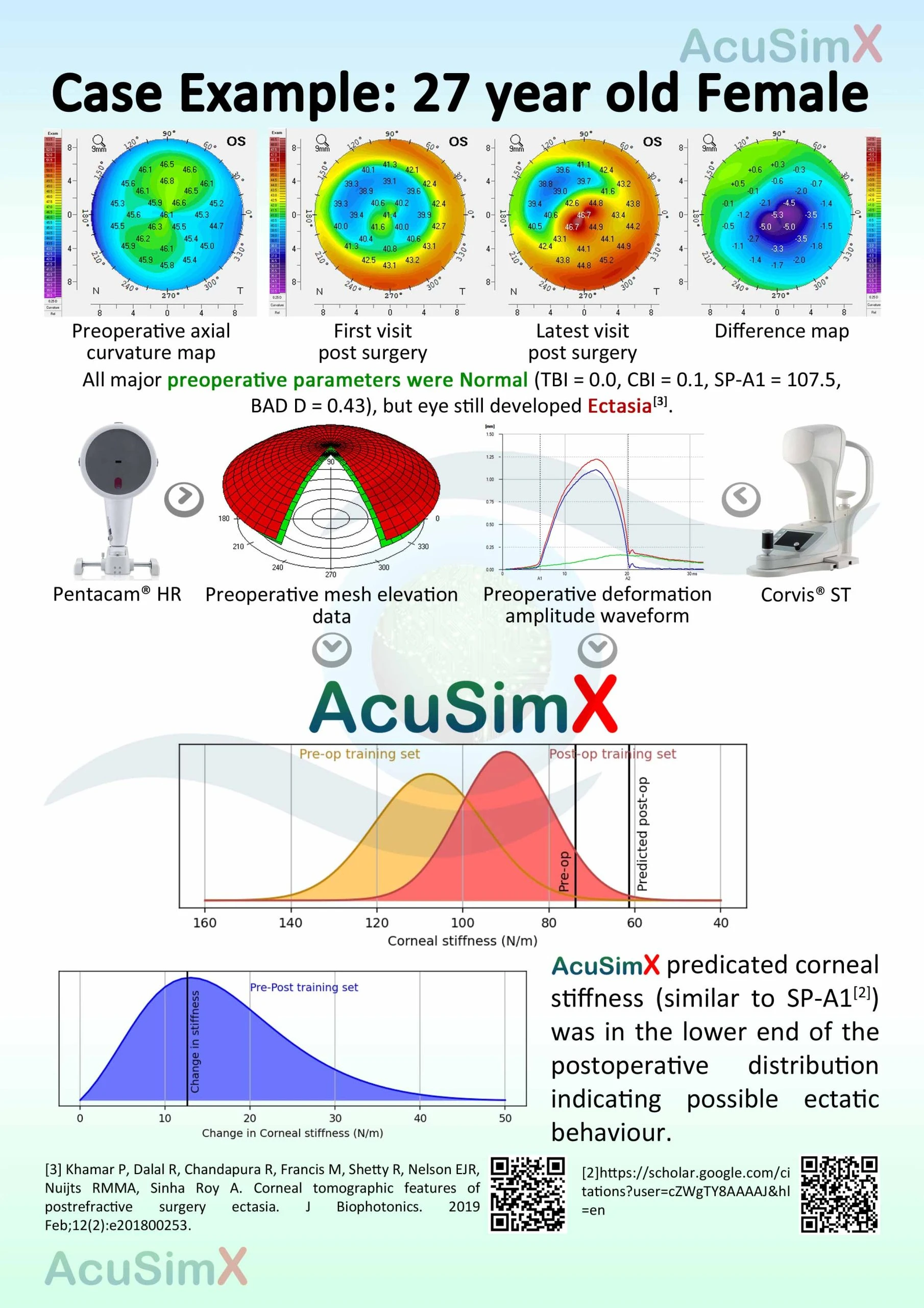
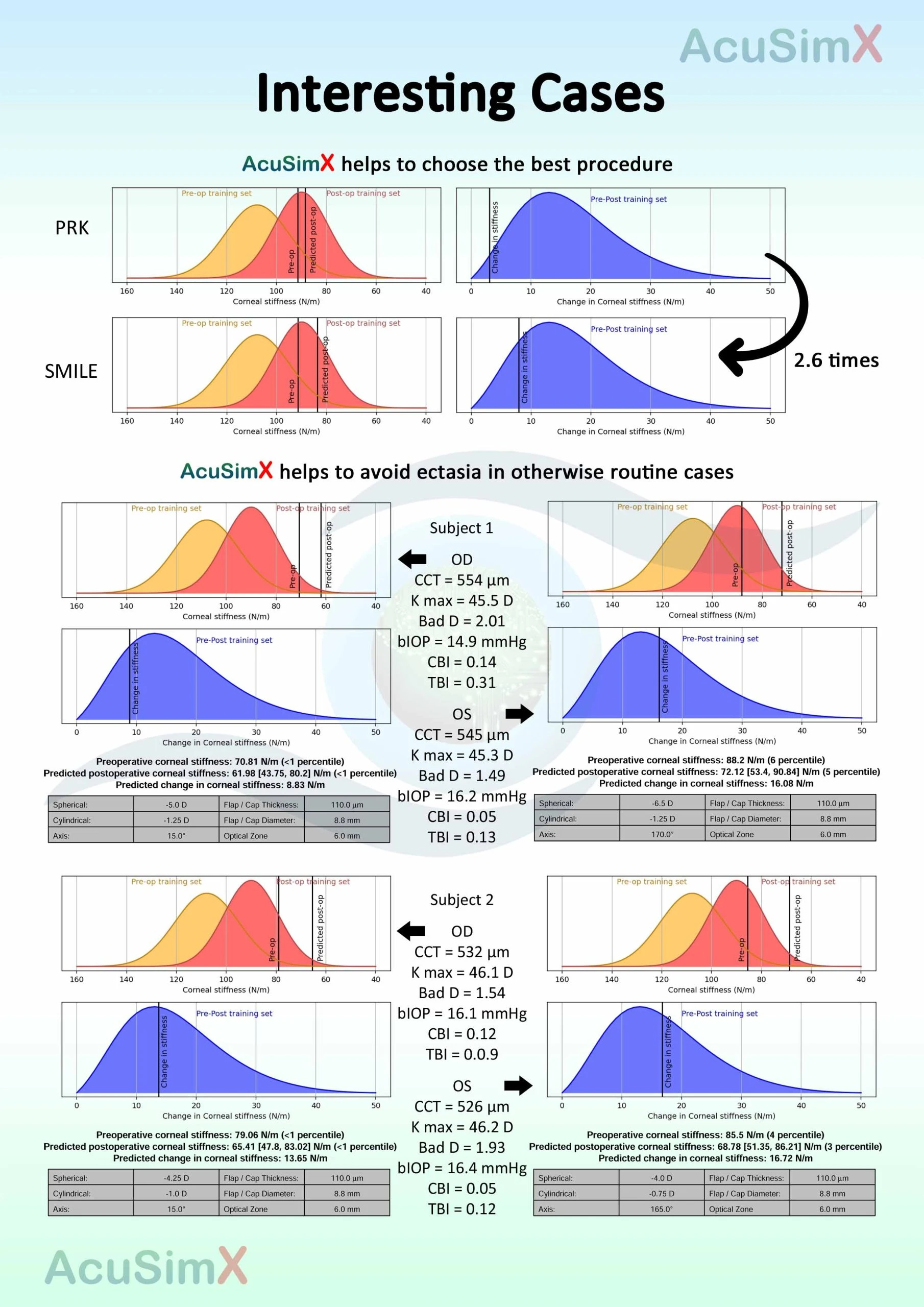
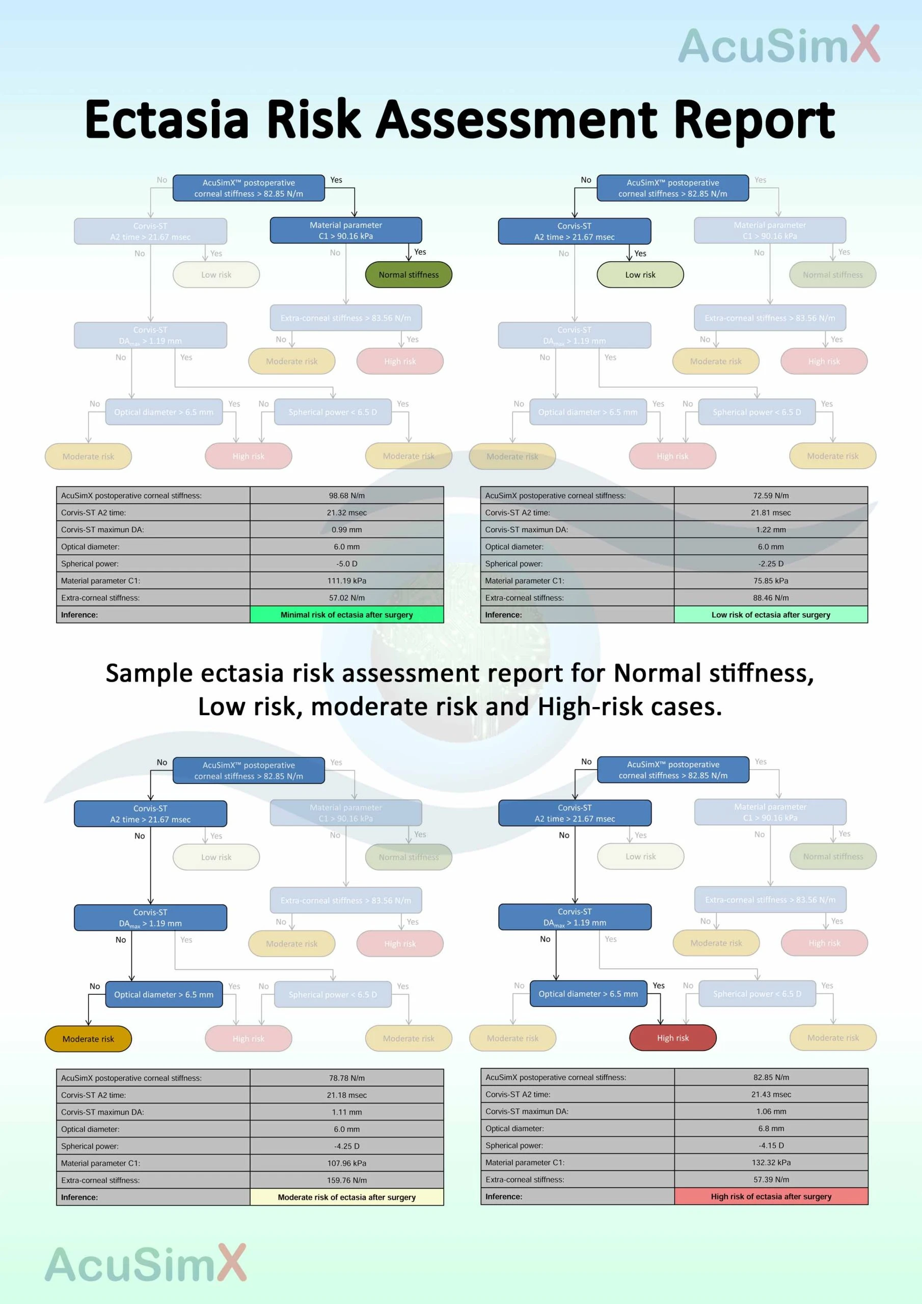
Laser refractive surgery is safe and effective, but it weakens corneal tissue by removing and reshaping it. If weakening exceeds a threshold, the cornea bulges outward, causing vision decline (ectasia), a critical risk with global surgery volumes projected at 5.8 million procedures. IBMS’s AcuSimX, an AI-powered predictive simulation, assesses corneal strength pre- and post-surgery, enabling virtual procedure refinement (like spacewalk planning on the ISS) and customized crosslinking for keratoconus.
Objectives
The Corneal Biomechanics programme aims to:
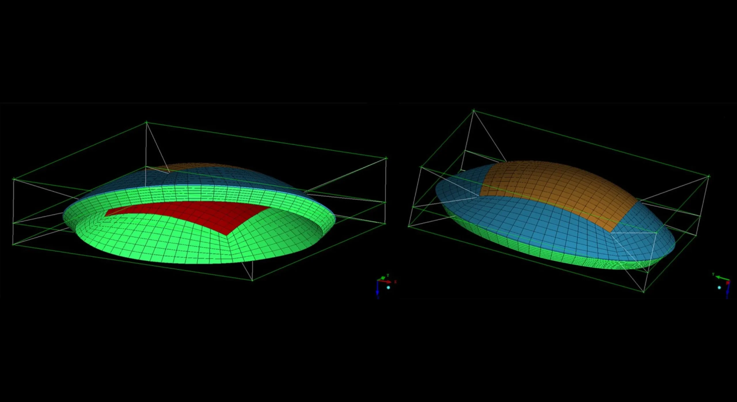
Quantify corneal mechanical behaviour using viscoelastic models and finite element analysis.
Predict risks such as post-surgery ectasia and keratoconus progression using AI tools like AcuSimX.
Improve surgical planning and outcome forecasting with patient-specific simulations.
Prevent complications and enhance visual quality through early intervention.
Core Research Focus
The corneal biomechanics programme focuses on understanding the mechanical behaviour of the cornea in health and disease.
Core areas of focus include:
Measurement and characterisation of corneal mechanical properties (strength and behaviour).
Biomechanical modelling of corneal structure, deformation, and tissue response.
Simulation-based approaches, including AI-driven AcuSimX for ectasia risk prediction.
Key translational focus areas include:
Early detection of biomechanical weaknesses in ectasia and keratoconus.
Development of diagnostic tools, like customized crosslinking protocols.
Integration of predictive simulations with surgical planning and clinical outcomes.
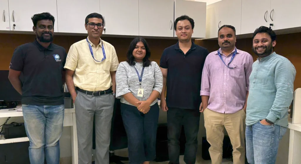
Together, these efforts support a diverse portfolio of Corneal Biomechanics research, with support from Engineers, Clinicians, and Imaging Scientists.
Team
The Corneal Biomechanics programme is led by a multidisciplinary team of scientists and clinician researchers with expertise in:
Engineers (viscoelastic and finite element modeling).
Clinicians (surgical and therapeutic outcomes).
Imaging Scientists (corneal assessment and simulation integration).
Close collaboration between laboratory researchers and clinicians ensures that research priorities remain aligned with patient needs and real-world clinical applicability.
