
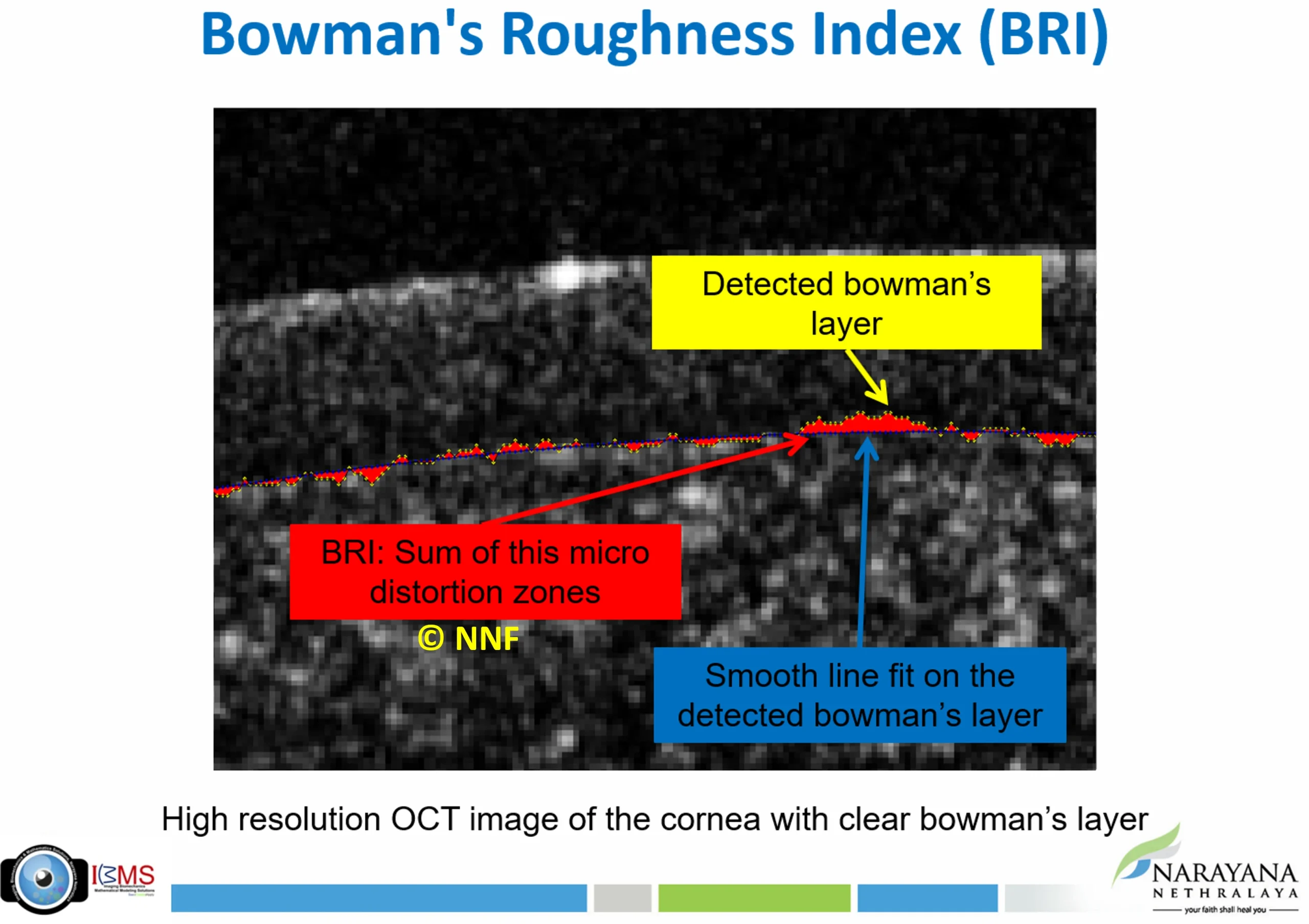
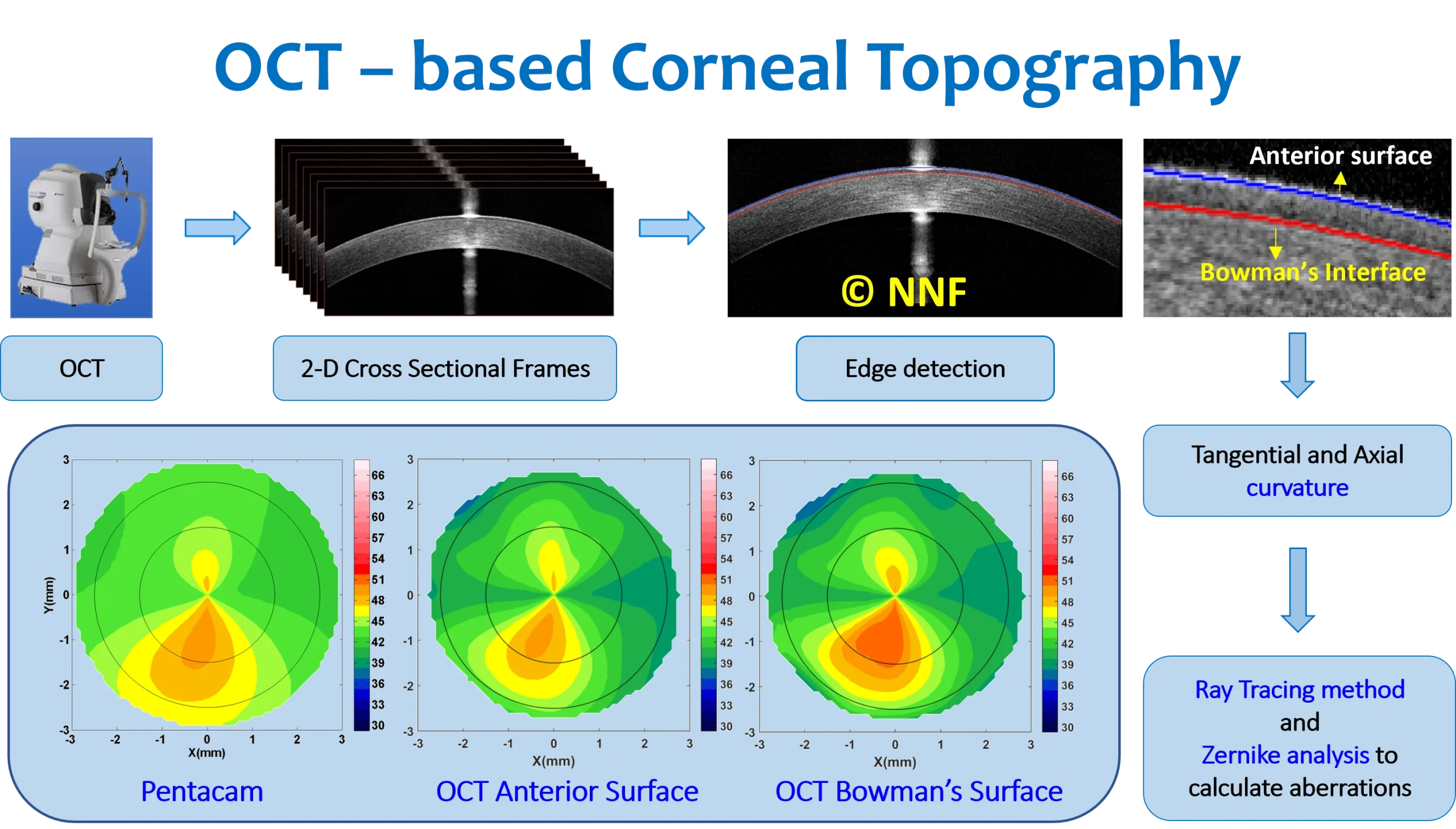
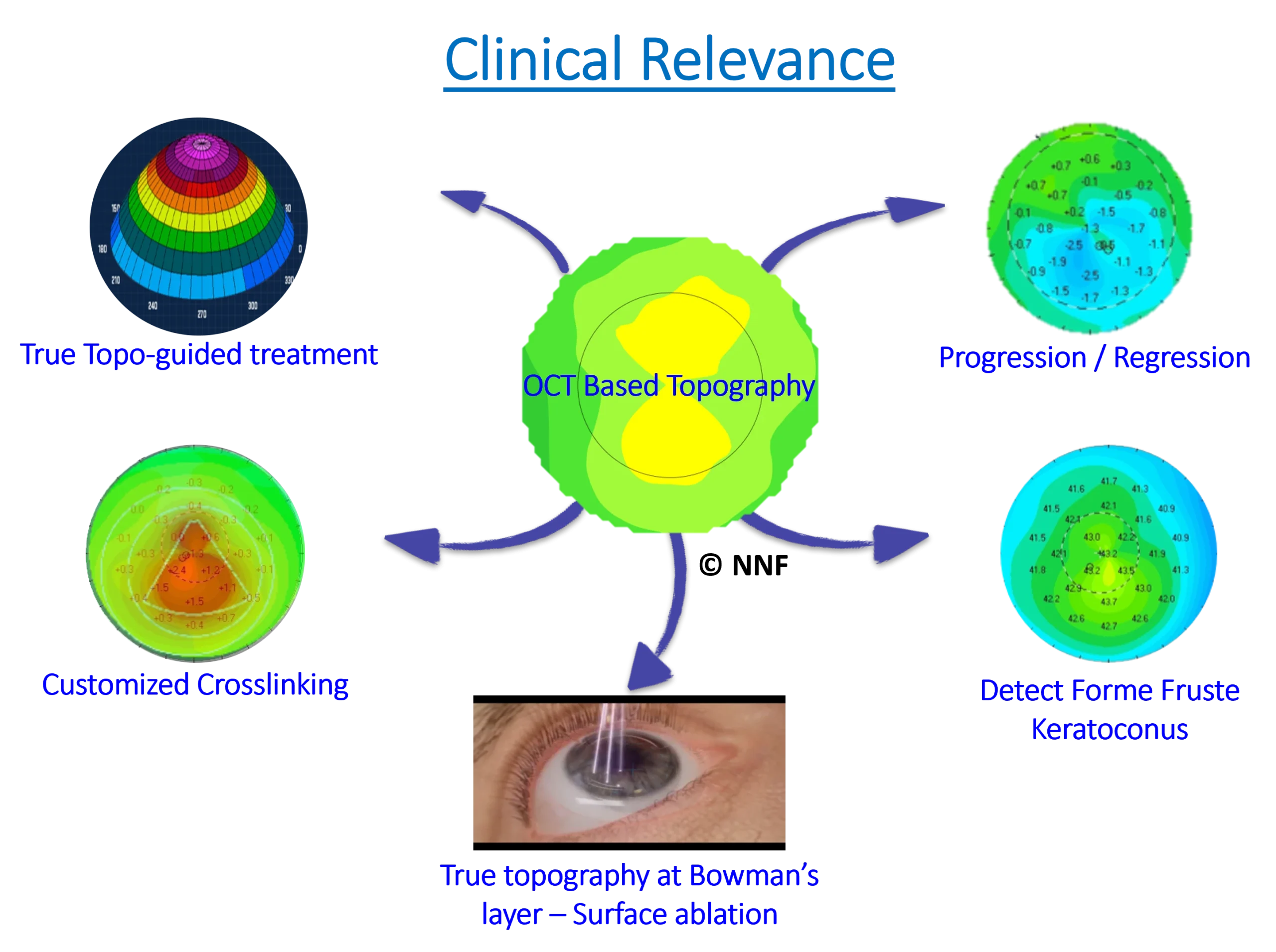
Optical coherence tomography (OCT) produces detailed cross-sectional images of the eye and is central to diagnosing conditions like keratoconus, glaucoma, macular degeneration, and diabetic retinopathy. IBMS develops next-generation imaging tools and automated algorithms for quantitative evaluation of both corneal and retinal OCT images. In corneal imaging, IBMS was among the first in the field to develop anterior corneal and Bowman’s layer topography and aberration maps from high-resolution OCT.
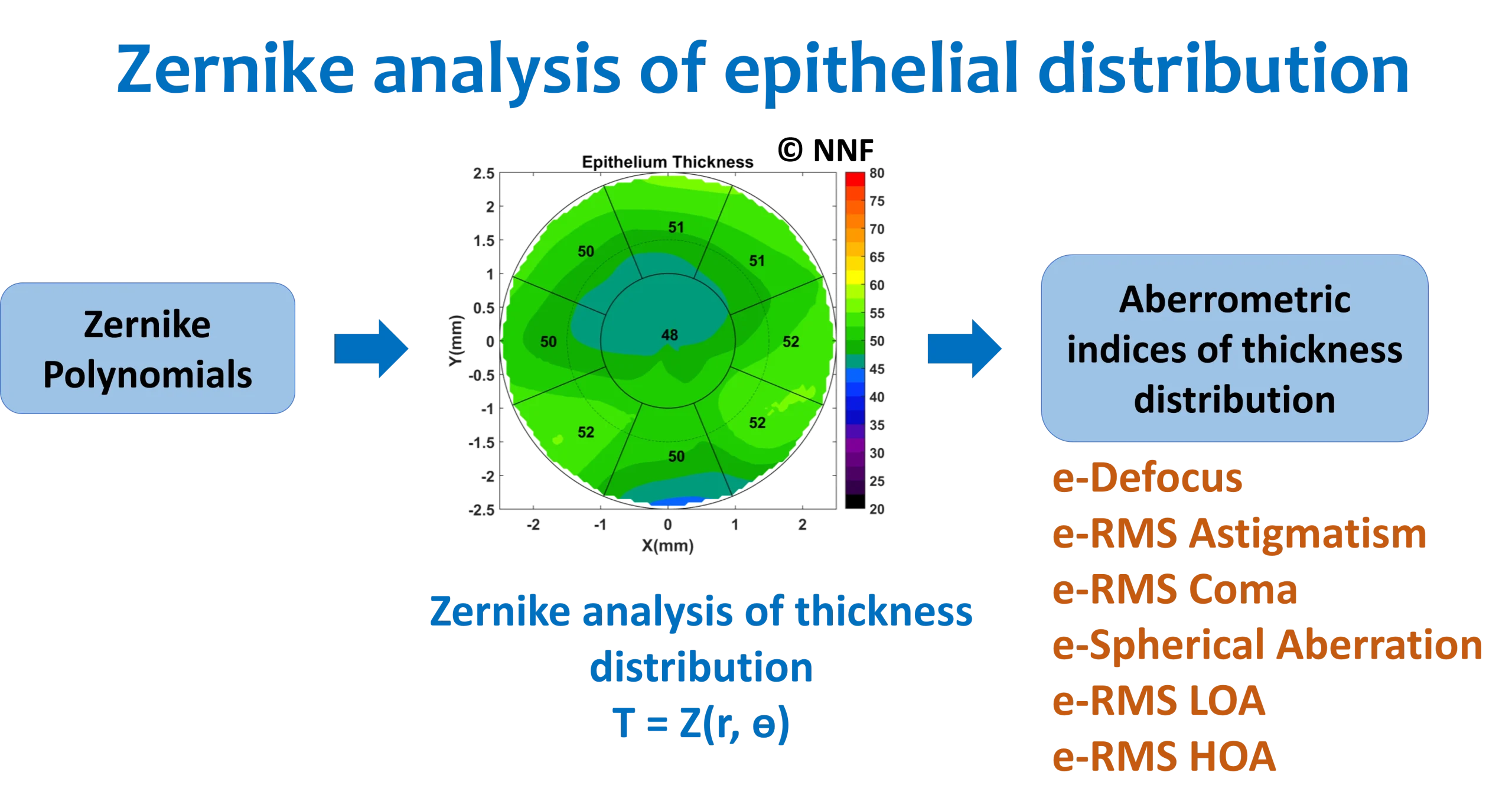
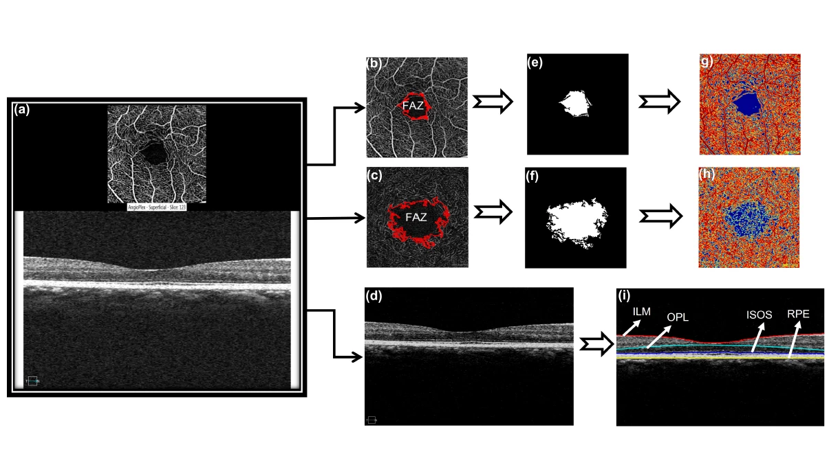
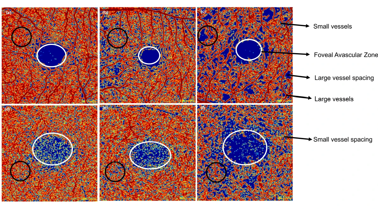
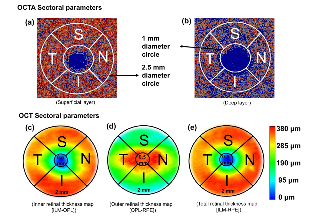
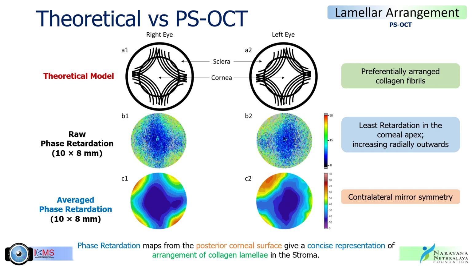
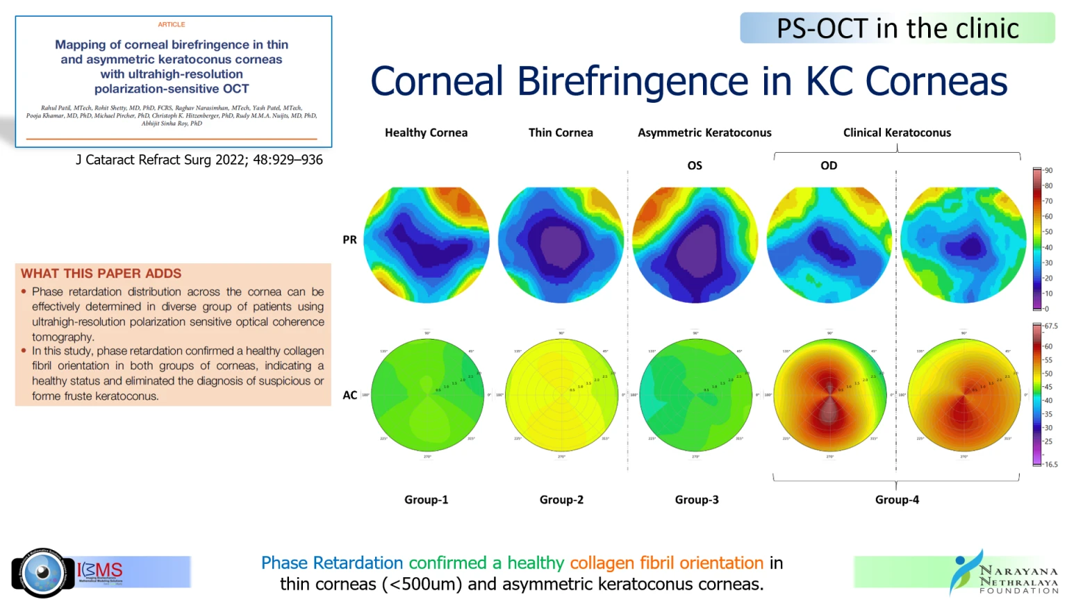
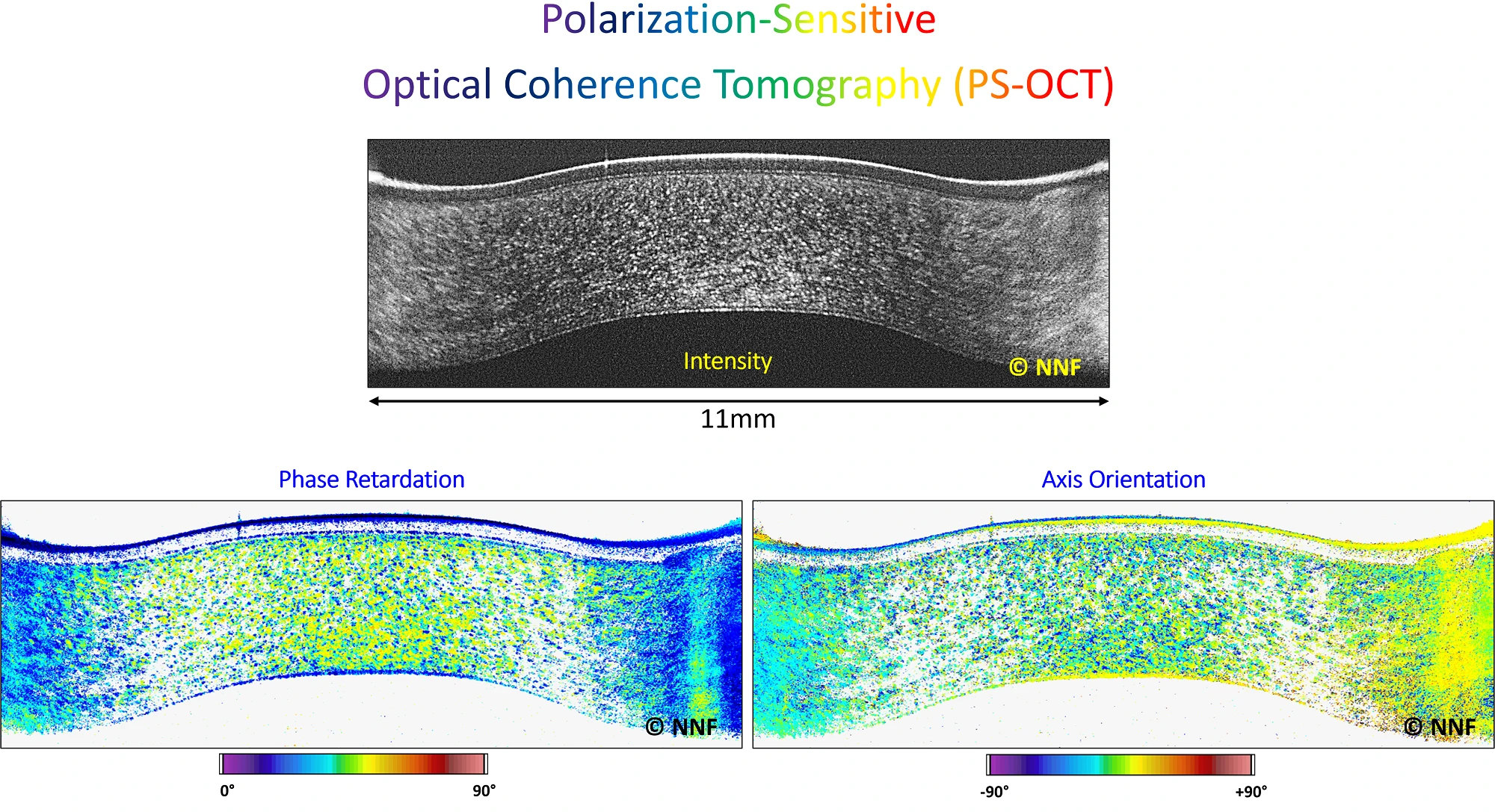
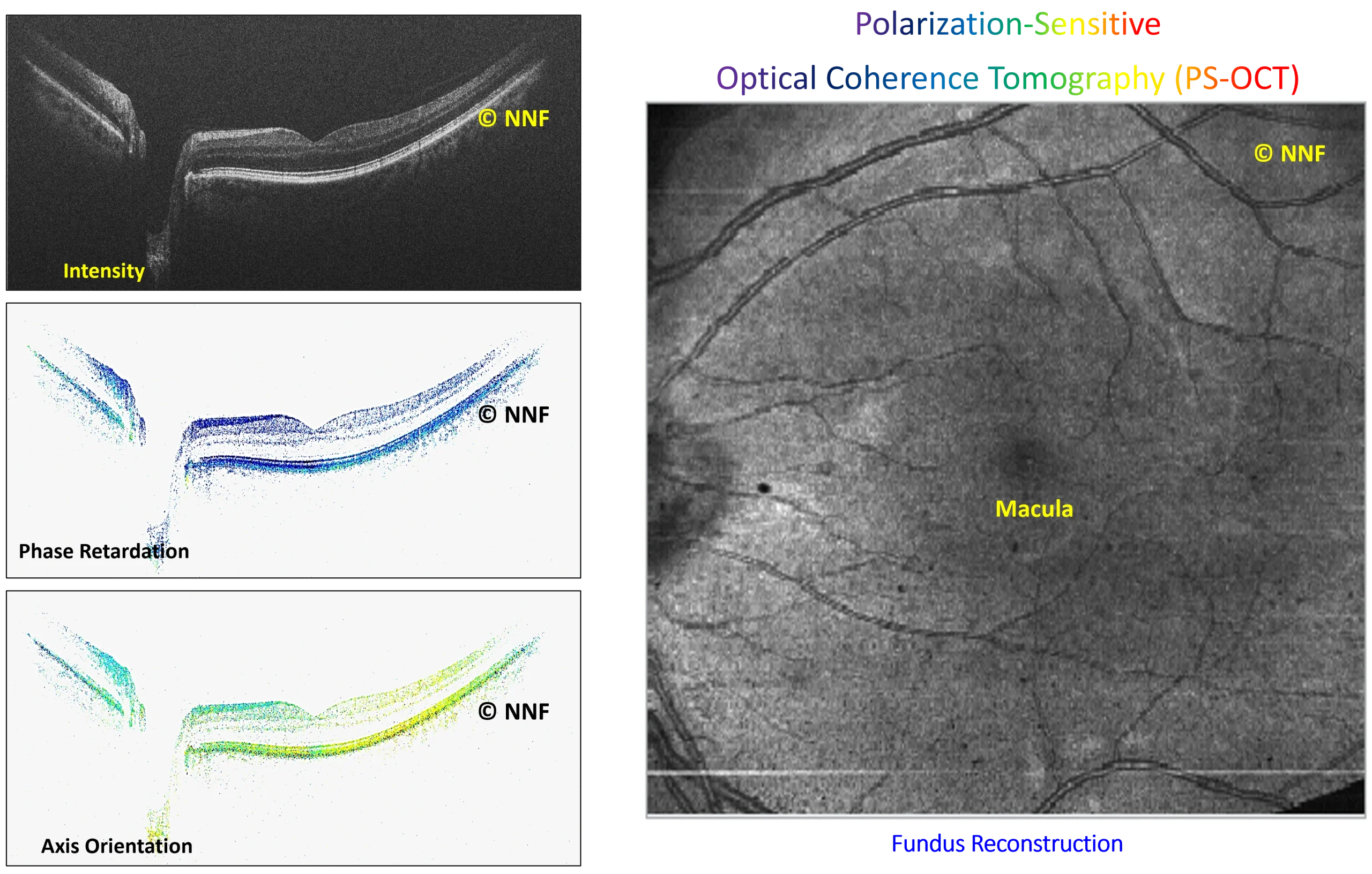
The lab has also serially evaluated epithelial remodelling following procedures such as LASIK, SMILE, and PRK, and quantified micro-distortions in the Bowman’s layer to support the identification of asymmetric keratoconus. In retinal imaging, the lab has developed automated algorithms to quantify OCT images in glaucoma and diabetic retinopathy, evaluated posterior vitreous visibility using enhanced depth and vitreous imaging techniques, and conducted extensive OCTA vascular comparisons across devices, finding significant correlations between macular edema volume and retinal features in patients with diabetes. IBMS also pioneers Polarization-Sensitive OCT (PS-OCT), which captures birefringence, a key biomarker for early disease detection, to reveal microstructural details invisible to conventional OCT. In parallel, IBMS is developing Cellular-Resolution OCT to visualise corneal structures at near-microscopic detail in real time, without contact or anaesthesia, bridging the gap between clinical imaging and laboratory microscopy.
The Advanced Ocular Imaging programme aims to:
The advanced ocular imaging programme focuses on quantitative evaluation of corneal and retinal structures.
Core areas of focus include:
Additional emphasis is placed on:
Together, these efforts support a diverse portfolio of Advanced Ocular Imaging research across multiple areas and engage both Imaging Scientists and Clinical Researchers.
The Advanced Ocular Imaging programme is led by a multidisciplinary team of scientists and clinician researchers with expertise in:
Close collaboration between laboratory researchers and clinicians ensures that research priorities remain aligned with patient needs and real-world clinical applicability.
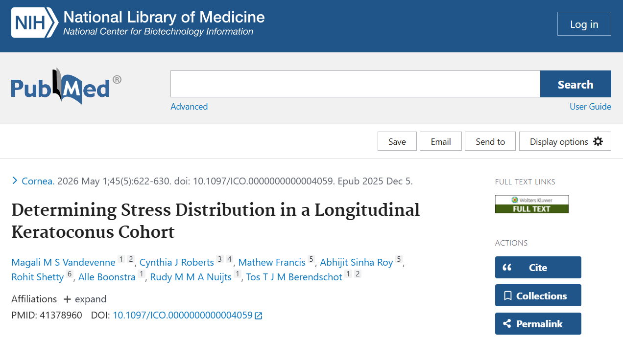
Authors: Vandevenne MMS, Roberts CJ, Francis M, Sinha Roy A, Shetty R, Boonstra A, Nuijts RMMA, Berendschot TTJM
Keywords: NA
PMID: 41378960
DOI: 10.1097/ICO.0000000000004059
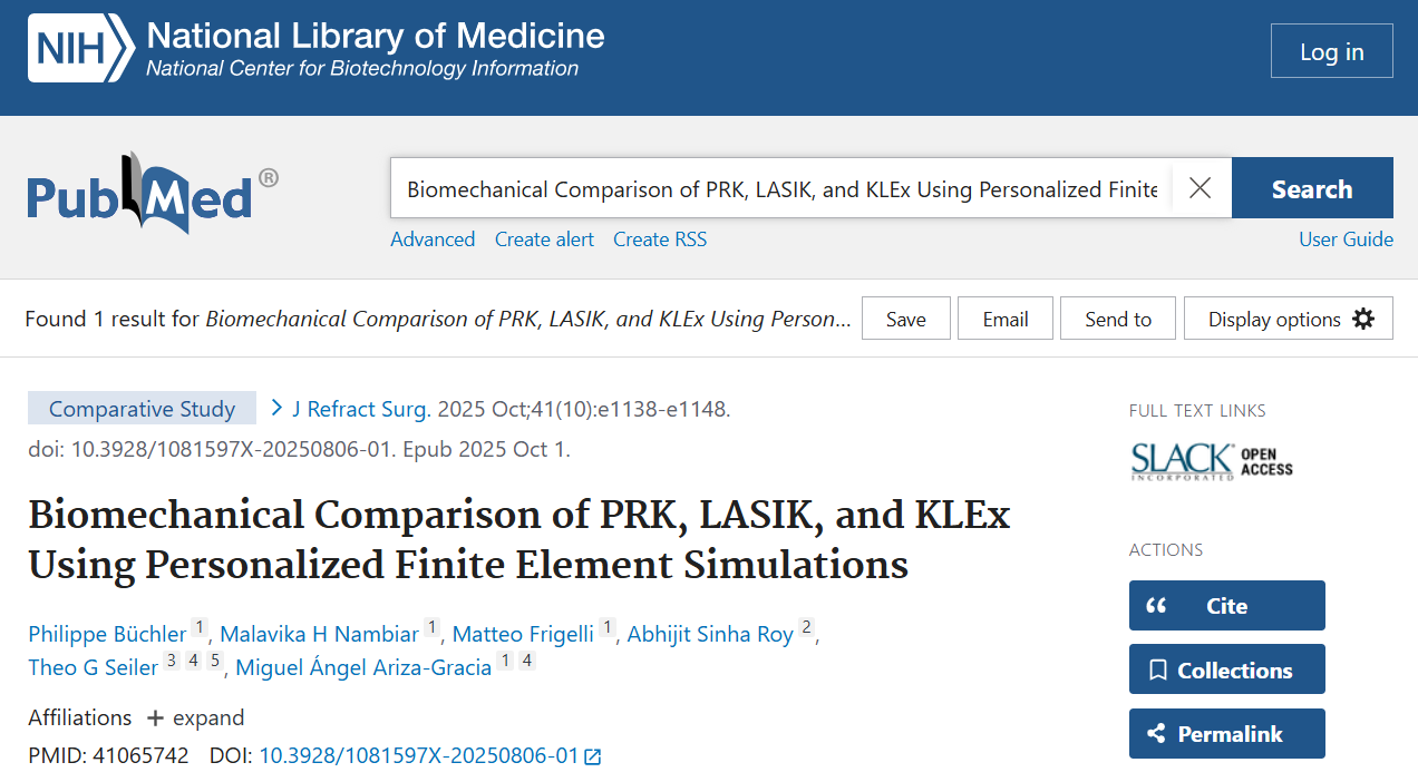
Authors: Büchler P, Nambiar MH, Frigelli M, Sinha Roy A, Seiler TG, Ariza-Gracia MÁ
Keywords: NA
PMID: 41065742
DOI: 10.3928/1081597X-20250806-01
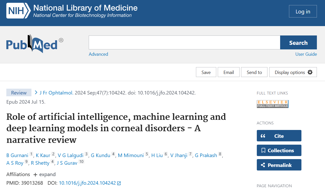
Authors: Gurnani B, Kaur K, Lalgudi VG, Kundu G, Mimouni M, Liu H, Jhanji V, Prakash G, Sinha Roy A, Shetty R, Gurav JS
Keywords: NA
PMID: 39013268
DOI: 10.1016/j.jfo.2024.104242

The PS-OCT and Cellular-Resolution OCT programme is progressing towards: