
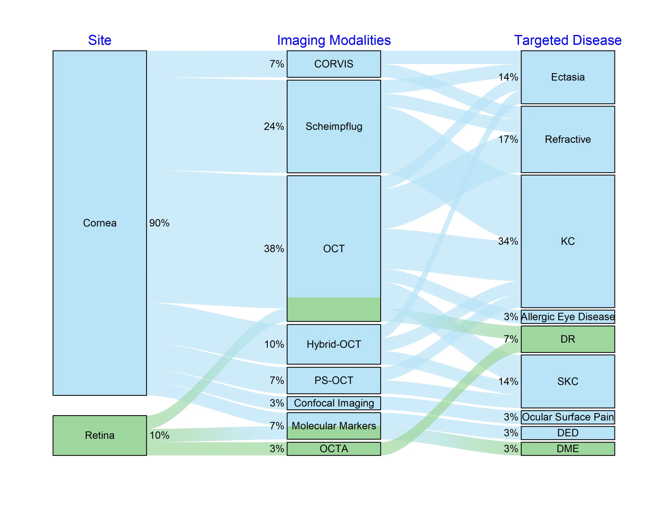
AI is transforming ophthalmology diagnostics and care. At IBMS, AI is applied across corneal and retinal imaging to surface findings that conventional methods miss and to accelerate clinical decision-making. In corneal work, the lab has used AI to identify vascular changes preceding tomographic changes in diabetic eyes, detect differential epithelial remodelling after LASIK, SMILE, and PRK, uncover unique corneal features of allergic eye disease, and combine corneal confocal microscopy with clinical features to evaluate ocular surface pain.
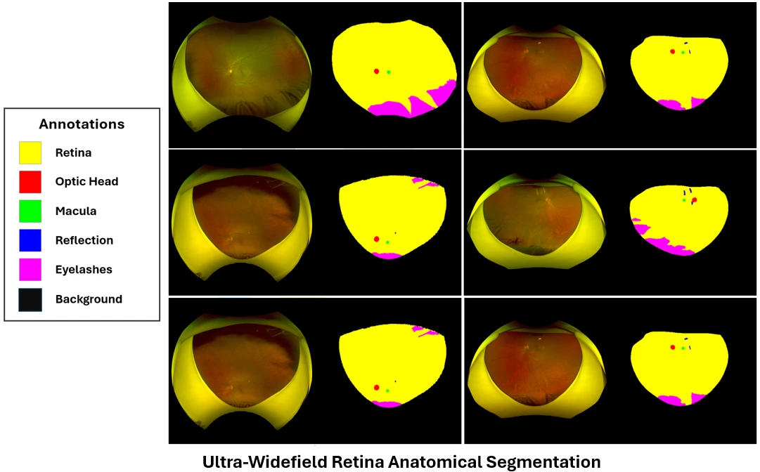
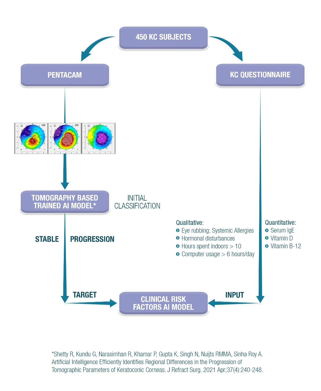
AI has also identified regional differences in tomographic parameters of keratoconic corneas and powered a universal architecture for early keratoconus diagnosis. The lab’s most recent work uses AI-based stratification of high-risk demographic and ocular-surface factors to study keratoconus progression. In retinal work, the lab builds intelligent systems that assist clinicians by analysing retinal images and highlighting important diagnostic patterns, with a focus on expanding access to specialist-level screening.
The Artificial Intelligence programme aims to:
The artificial intelligence programme focuses on understanding AI applications in ophthalmology diagnostics and care.
Core areas of focus include:
Key technical directions include:
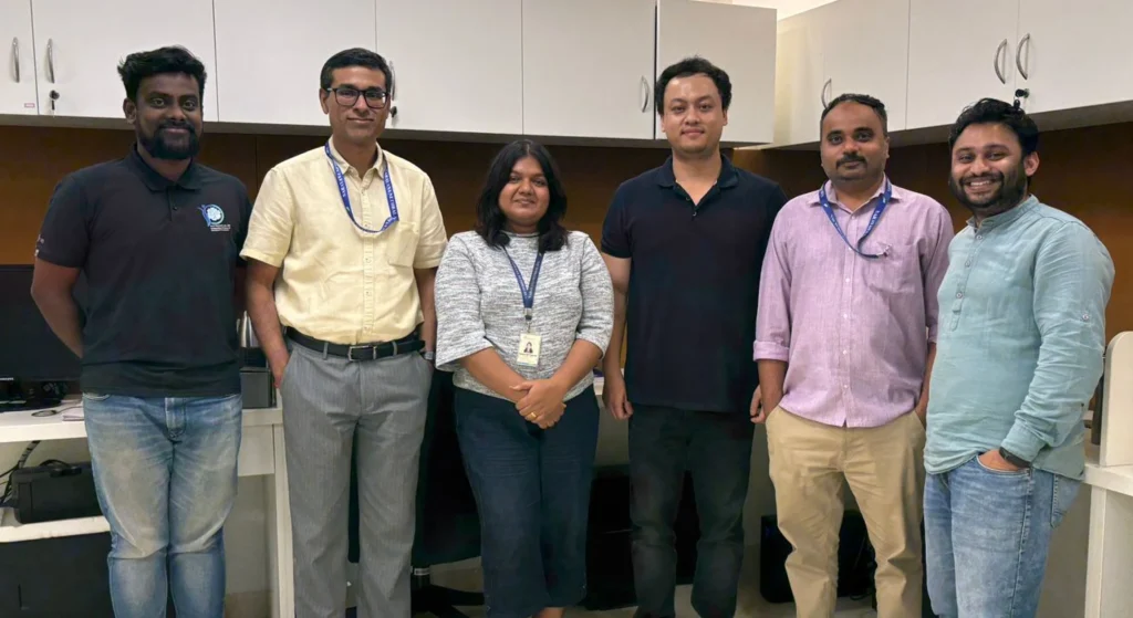
Together, these efforts support a diverse portfolio of Artificial Intelligence research, with support from Engineers, Clinicians, and Imaging Scientists.
The Artificial Intelligence programme is led by a multidisciplinary team of scientists and clinician researchers with expertise in:
Close collaboration between laboratory researchers and clinicians ensures that research priorities remain aligned with patient needs and real-world clinical applicability.
Authors: Vandevenne MMS, Roberts CJ, Francis M, Sinha Roy A, Shetty R, Boonstra A, Nuijts RMMA, Berendschot TTJM
Keywords: NA
PMID: 41378960
DOI: 10.1097/ICO.0000000000004059
Authors: Büchler P, Nambiar MH, Frigelli M, Sinha Roy A, Seiler TG, Ariza-Gracia MÁ
Keywords: NA
PMID: 41065742
DOI: 10.3928/1081597X-20250806-01
Authors: Gurnani B, Kaur K, Lalgudi VG, Kundu G, Mimouni M, Liu H, Jhanji V, Prakash G, Sinha Roy A, Shetty R, Gurav JS
Keywords: NA
PMID: 39013268
DOI: 10.1016/j.jfo.2024.104242
The AI and Retinal Imaging programme is progressing towards: